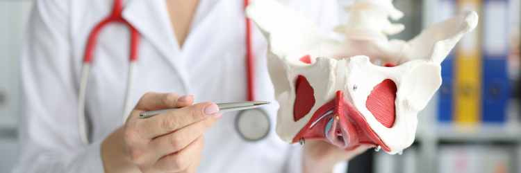
I love how we use technology to attack a growing problem—especially in medicine. This one has particular relevance for me. I went from having the bones of a 35-year-old to osteoporosis in two years due to a medication regimen. I also fell off a deck and fractured my “sitting bone” (not tailbone) in two places. The fall was due to a large and crazy dog!
While the risk of bone fractures due to osteoporosis increases with age, the medical community is considering 50 and older “aged.” Annually, approximately 1.5 million people suffer a fracture because of bone disease. In fact, fractures may be the first visible sign of the disease. In the U.S., over 10 million people aged 50 and above have osteoporosis of the hip. 33.6 million have low bone mass or “osteopenia” of the hip and are at risk for osteoporosis.
Currently, diagnosis for osteoporosis involves bone density measurement – a 2D X-ray examination that is typically first used once a fracture occurs. Bone density tests are not reliable. According to Hanna Isaksson, professor of biomedical engineering at Lund University’s Faculty of Engineering (LTH), approximately half the people in the risk zone are missed.
A greater percentage of those with a high risk of sustaining a hip fracture could be identified using a new calculation and simulation method developed by Isaksson and her research team. The results of a study evaluating their process involving 2,000 people was published in the Journal of Bone and Mineral Research.
The new method uses 2D X-ray images from bone density measurements to create 3D models of the thigh bone. 2D to 3D uses a computer-simulated template that describes how the bone’s geometry and bone density vary. The 3D model of the thigh bone enables the simulation of different situations and scenarios that may have an effect. When results from both methods were compared, the 3D method is better at predicting the risk of a hip fracture than the bone density measurement.
The researchers claim they could identify around 1,000 more people yearly with a high risk of hip fracture. The method can also be used to identify osteoporosis before the first fracture occurs.
More accurate diagnosis could also reduce the number of deaths; for those who suffer a hip fracture, 24% die within one year.

