These 3D-Printed Microfish Were Created To Sniff Out And Remove Toxins
Swarms of microscopic fish are being created at the University of California San Diego.
Nanoengineers have developed a technique for creating 3D-printed robotic, microscopic fish that can swim in water and are powered by hydrogen peroxide.
The magnetically controlled microfish are being created in an attempt to develop a new class of “smart” microrobots that can perform tasks such as detoxification, sensing and directed drug delivery, according to a news release.
Usually microrobots are incapable of performing complex tasks like these due to their simplistic structure and design, but Professors Shaochen Chen and Joseph Wang of the NanoEngineering Department at the UC San Diego have now demonstrated a new method for creating microrobots that allows the robots to do more.
To do this, the team of nanoengineers added functional nanoparticles into parts of the microfish bodies. Then they installed platinum nanoparticles into the tails (doing so causes a reaction with hydrogen peroxide and propels the microfish forward) and injected magnetic iron oxide nanoparticles into the heads (this allowed them to be steered by the magnets).

In order to create the microfish bodies, the researchers employed a production method called microscale continuous optical printing (μCOP), developed by Chen, which offers benefits such as speed, scalability, precision, and flexibility. In just seconds they were able to print an array containing hundreds of microfish, each measuring 120 microns long and 30 microns thick.
“We have developed an entirely new method to engineer nature-inspired microscopic swimmers that have complex geometric structures and are smaller than the width of a human hair. With this method, we can easily integrate different functions inside these tiny robotic swimmers for a broad spectrum of applications,” said Wei Zhu, a nanoengineering Ph.D. student in the research group.
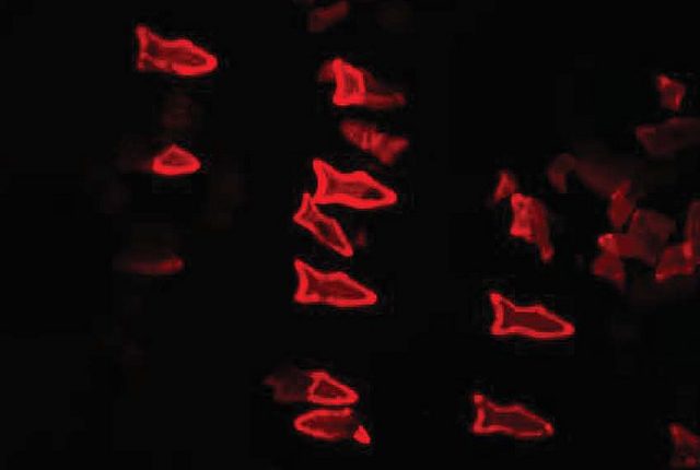
To demonstrate the technology, the team placed toxin-neutralizing nanoparticles in the bodies of the microfish (in this case they used polydiacetylene (PDA) nanoparticles which captures harmful pore-forming toxins like the kind found in bee venom). When the microfish were placed in the toxic water, the PDA nanoparticles stuck to the toxin molecules and lit up in a fluorescent red light. The team was able to determine how well the microfish can clean up toxins by the intensity of their red glow.
“Another exciting possibility we could explore is to encapsulate medicines inside the microfish and use them for directed drug delivery,” said Jinxing Li,a nanoengineering Ph.D. student on the team.
To learn more about the process visit the University of California, San Diego website.

