New approach to microfluidics for quicker detection of CTCs
Circulating Tumor Cells (CTC) are key early indicators of metastasis, which is the process by which cancer cells move from one organ group in the body to another. Once cancer spreads, the prognosis is generally not good. So, early identification of CTCs can help prevent them from creating new colonies of malignant cells.
Researchers at Worcester Polytechnic Institute (WPI) in Massachusetts have developed a new approach to microfluidics to detect CTCs in blood. The WPI researchers believe that their technique could form the basis of a simple lab test for quick detection of early signs of metastasis and help physicians select treatments targeted at the specific cancer cells identified.
Current microfluidic techniques used in tumor cell isolation have been dependent on flow rate and require off-chip post-processing. The WPI researchers’ technique employs static isolation of tumor cells from the blood by fractionation of the blood into small droplets.
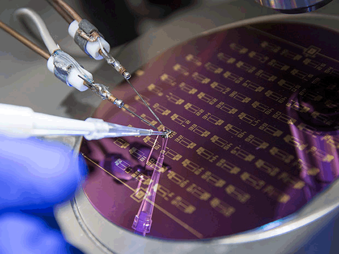
In research described in the journal Nanotechnology, the WPI researchers were able to create a chip design in which antibodies are attached to an array of carbon nanotubes at the bottom of a tiny well in the chip. The chips have an array of these tiny wells, each about three millimeters across.
When the blood droplets are put into the well, the heavier cancer cells drop to the bottom where they become attached to the antibodies. Each of the wells holds a specific antibody that will bind to one type of cancer cell. The chip’s electrodes detect electrical changes that occur when the cancer cells are captured by the antibodies.
Using an array of antibodies makes it possible to identify several different types of cancer cells within a single blood sample. To put that in perspective, the researchers could fill 170 wells with just 0.85 millileter of blood. The chips were able to capture between one and a thousand cells per device, equating to an efficiency of between 62 and 100%.
You can see a video that offers a demonstration of how the chip works below.
The advantages of this technique over traditional microfluidic methods are numerous and significant. But let’s just focus on the advantages derived from the use of carbon nanotubes.
First, the nanotube-based microarrays include both detection and capture technology, unlike traditional microfluidics, which only capture. Second, the nanotube microarray allows for a wide variety of antibodies so that it can attract and identify different types of cells that may need to be fought in different ways.
Another one of the advantages of this approach over other microfluidics is that it can capture exosomes, which are produced by cancer cells and carry the same markers.
“These highly elusive 3-nanometer structures are too small to be captured with other types of liquid biopsy devices, such as microfluidics, due to shear forces that can potentially destroy them,” said Balaji Panchapakesan, associate professor of mechanical engineering at WPI and director of the Small Systems Laboratory, in a press release. “Our chip is currently the only device that can potentially capture circulating tumor cells and exosomes directly on the chip, which should increase its ability to detect metastasis.” Panchapakesan adds that this is important because research is showing that tiny proteins excreted with exosomes can actually suppress cancer drug delivery and hinder treatment.
Panchapakesan believes the technology is ready for commercialization, but his team just needs more data on patients (delineated by stage of cancer) to move the technology along further in its development.
In an e-mail interview with IEEE Spectrum, Panchapakesan added: “If there is any equipment that needs to be developed more, [it’ll probably be] the automation and robotic handling of the entire system from drop deposition to microscopy. But really we just need patients, patients, patients.”
More information: IEEE Spectrum


Comments are closed, but trackbacks and pingbacks are open.